The first paper in Brain Pathology
Abstract
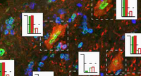
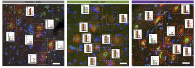
Histopathological analysis of tissue sections is invaluable in neurodegeneration research. However, cell‐to‐cell variation in both the presence and severity of a given phenotype is a key limitation of this approach, reducing the signal to noise ratio and leaving unresolved the potential of single‐cell scoring for a given disease attribute. Here, we tested different machine learning methods to analyse high‐content microscopy measurements of hundreds of motor neurons (MNs) from amyotrophic lateral sclerosis (ALS) post‐mortem tissue sections. Furthermore, we automated the identification of phenotypically distinct MN subpopulations in VCP‐ and SOD1‐mutant transgenic mice, revealing common morphological cellular phenotypes. Additionally we established scoring metrics to rank cells and tissue samples for both disease probability and severity. By adapting this paradigm to human post‐mortem tissue, we validated our core finding that morphological descriptors robustly discriminate ALS from control healthy tissue at single cell resolution. Determining disease presence, severity and unbiased phenotypes at single cell resolution might prove transformational in our understanding of ALS and neurodegeneration more broadly.
Publication: Automated and unbiased discrimination of ALS from control tissue at single cell resolution, https://doi.org/10.1111/bpa.12937

